Foreword
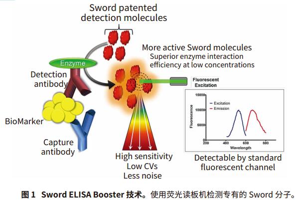
Traditional ELISA (Enzyme-Linked Immunosorbent Assay) method using horseradish peroxidase (HRP) substrate 3, 3', 5, 5'-tetramethylbenzidine (TMB) is generally unable to detect low abundance analytes, For example, inflammatory cytokines. SwordDiagnostics has developed a new generation of ELISA detection technology, the Sword ELISA Booster, which can directly replace the traditional ELISA detection reagents as a gold standard, increasing sensitivity by 30 times.
Sword ELISA Booster performs immunochemical assays by adding proprietary detection reagents. The Sword molecule is converted to a resonant Raman-active substrate in the presence of horseradish peroxidase (HRP) and the resonant Raman signal is detected on a microplate reader. Due to the extremely high level of enzyme/substrate interaction at low concentrations, more resonant Raman-active molecules are produced, enhancing the performance of the experiment. This experiment has higher sensitivity, lower CV, and less noise than traditional ELISA assay reagents.
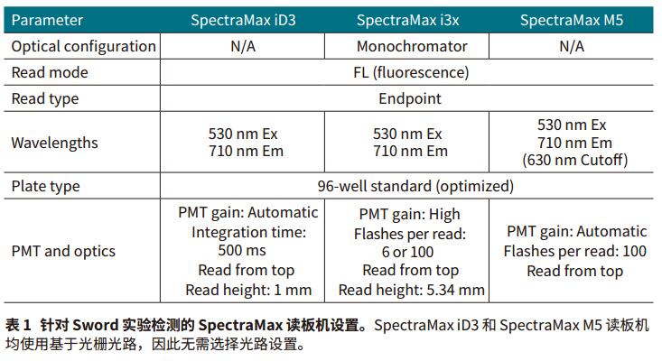
Here, we used the Sword ELISA Boosters assay to detect the inflammatory cytokines TNF-α and IL-1β in the fluorescence mode of Molecular Devices' SpectraMax® iD3, i3x and M5 multifunctional microplate readers. The two analytes having a concentration as low as pg/mL were respectively detected on the above three plate readers.
advantage
- High sensitivity detection of low abundance analytes
- Enhancement of traditional ELISA reagents by proprietary Sword molecules
- Low background noise and CV values
material
- Sword ELISA Booster for human IL-1β detection (Sword Diagnostics cat.#SB-HIL1B02-05)
- Human IL-1β/IL-1F2 DuoSet ELISA (R&DSystems cat. #DY201)
- Sword ELISA Booster for human TNF-α detection (Sword Diagnostics cat.#SB-HTNFA02-05)
- Human TNF-α DuoSet ELISA (R&D Systemscat. #DY210)
- An immuno-cleaning standard module (striped plate) coated with Nunc MaxiSorp (Thermo Scientific cat. #445101)
- SpectraMax iD3 multi-function microplate reader
- SpectraMax i3x Multi-Purpose Microplate Reader
- SpectraMax M5 Multi-Purpose Microplate Reader
- MultiWash+ washing machine
method
Prepare the working fluid and the orifice plate. The experimental procedure of the two kits is as follows. Please refer to the product manual for more details on reagent handling and methods.
Sword ELISA Booster substrate solution
To prepare 16 mL of Sword ELISA Booster substrate solution sufficient for 96-well plates, add 11.2 mL of deionized water to the following reagents:
- 1.6 mL Sword Booster Component A (10x)
- 1.6 mL Sword Booster Component B (10x)
- 1.6 mL Sword Booster Component C (10x)
1x Sword Development solution
To prepare 16 mL of Sword Development solution for 96-well plates, add 3.2 mL of 5x Sword Development Solution (Component D) to 12.8 mL of deionized water.
Prepare the orifice plate
IL-1β and TNF-α capture antibodies were redissolved in 0.5 mL PBS to prepare antibody concentrates. The concentrated human TNF-α capture antibody was diluted to 12 μg/mL in PBS, and the human IL-1β capture antibody was diluted to 4 μg/mL to prepare a working concentration. Each experimental well plate was coated with 100 μL/well of diluted antibody. The plates were then sealed and incubated overnight at 4 °C.
The plate was washed 3 times with 400 μL/well wash buffer (PBS + 0.5% Tween20) using a MultiWash+ microporous plate washer, leaving the wash buffer in the well for 15-30 seconds before each pipetting. Each plate was then blocked with 200 μL/well of Sword ELISA Blocker for IL-1β or TNF-α, sealed and incubated for at least 1 hour at room temperature. The orifice plate was washed again as described above.
Measurement process
result
Get the full content click the button below

Urinalysis test strips refer to test strips that test for bilirubin, urobilinogen, ketone bodies, ascorbic acid, glucose, protein (albumin), blood cells, PH, etc. in urine.
Detection principle
1. pH: The pH value in the range of 5-9 is measured by the pH indicator, and the pH value of the fresh urine of a normal person is between 5-7.
2. Nitrite: The reaction is based on the reduction of nitrate to nitrite by Gram-positive bacteria in the urine. The nitrite reacts with p-aminobenzenesulfonic acid to form diazonium compounds, which are then combined with N-(1-naphthalene) )-3 aminopropanesulfonate combined with a pink color.
3. Glucose: According to the reaction principle of glucose oxidase, glucose oxidase specifically oxidizes glucose to generate glucuronic acid and hydrogen peroxide. Under the action of hydrogen peroxide, hydrogen peroxide oxidizes the indicator and turns color. .
Classification
Urinalysis test strips are divided into visual series and machine series. The visual inspection series is divided into several models according to different inspection items; the machine inspection series is divided into several models according to different applicable instruments.
1. Classification by measurement method
1) Visual inspection series
When observing the result, compare the color with the standard color code within the time specified on the color code, judge and read the result.
2) Machine test series.
For instrument operation, refer to the instruction manual of the Urine Analyzer used.
2. According to the number of measurement items
There are single-item, 2-item, 4-item and multiple test strips. Currently, 10-item or 11-item multiple test strips are most commonly used in hospitals.
3. Classification by structure
Urinalysis test strips with single-layer membrane structure and multi-layer membrane structure.
Urine Reagent Strips,Urine Test Strip,Urine Sugar Strip Test,Visual Urine Analysis Strips
Jilin Sinoscience Technology Co. LTD , https://www.jilinsinoscience.com