Microcarrier ball transfer amplification of Vero cells in a WAVE reactor
Lu Lifang, Christain Kaisermayer, Yao Wei, Yan Lili
General Electric Medical Group Life Sciences, Fast Trak R&D Center, Shanghai
summary
Vero cells can be widely used in the production of vaccines. The successful amplification of Vero cell culture technology is critical to the large-scale application of this technology to vaccine production. As adherent cells, our experience has shown that Vero cells can successfully grow in WAVE reactors using microcarrier technology. To further explore the possibility of scale-up culture, we performed a ball-to-ball experiment on the microcarrier Cytodex 1 in a WAVE reactor. A series of experiments have been quite successful. The Vero cells were first cultured in a 10 L culture bag with Cytodex 1 microcarriers to a certain density, and the cells grown on the microcarriers were subsequently digested with trypsin, and the cells which were substantially detached from the microcarriers after digestion were finally and the old microcarriers were The passaging density was transferred to a new microcarrier culture system and a new round of cell culture was initiated. Our results show that the growth rate of cells is basically the same before and after several such transfer passages. It is entirely possible that the microcarrier culture technique of Vero cells is amplified in the WAVE reactor. Further experiments have shown that under the current working conditions, Vero cells can be up to 10 times magnification on Cytodex and ball transfer. This provides reliable support for new prospects for large-scale industrial production of cell culture-based vaccines.
Foreword
Vero cells originally originated from African green monkey kidneys and are known to be used against a variety of viruses such as SV40, SV-5, measles virus, arbovirus, respiratory enterovirus, rubella, sputum adenovirus, poliovirus, influenza virus, deputy Influenza viruses, vaccinia viruses, etc. are all sensitive. Vero cells are thus widely used for vaccine development for the corresponding diseases [1]. Vero cells have recently been used in the development of influenza vaccines [2]. The well-known pharmaceutical company Baxter Healthcare successfully used Cytodex 3 microcarriers for the production of Vero cell flu vaccine, using a three-stage fermenter for microcarrier ball transfer amplification, and finally to the 6000L reactor culture scale. Novartis has also licensed cell vaccine production technology in its European cell culture plants. Novartis recently reached an agreement with the US Department of Health and Human Services (HHS), which sponsored the former $486 million to establish a mammalian cell vaccine production facility in North Carolina and plans to enter it in 2012 [3]. Cell vaccine technology, with its rapid and reliable characteristics, as a global trend, is expected to gradually replace the current chicken embryo vaccine technology.
As adherent cells, Vero cells need to be grown on a certain surface, and can be cultured in a small-sized state using a T-shaped flask or a roller bottle. Domestic microcarrier cell culture levels are quite different from those in foreign countries. Most of them are still inoculated directly into a 30L fermentor using a spinner/cell factory, and multiple 30L fermentors are used for parallel amplification. The rotary bottle culture is labor-intensive operation, and it is impossible to precisely control the parameters such as pH and dissolved oxygen, and has a high risk of contamination. The parallel enlargement of multiple small fermenters cannot avoid the difference between each reactor, increase the control point of the production process, is not conducive to the stability of product quality and the verification of the entire production process, and limits the production capacity.
Therefore, it is necessary to develop large-scale microcarrier cell culture technology to meet the needs of biotechnology development, and the microcarrier ball-to-ball amplification process is one of the key points [4, 5] for cultivation and production on a larger industrial scale. . Measuring the success of this technology, on the one hand, is the success of the transfer, and the other important aspect is whether the cells can have the same growth state after the transfer.
method
Culture of Vero cells in WAVETM reactor
In a 10 liter cell culture bag, Vero was adherently cultured on a microcarrier Cytodex 1 (Cyt. 1) at a microcarrier concentration of 3-6 g/L and a suspension culture volume of 1.5-3 L. On the day before inoculation, the cell culture bag was pre-placed on a WAVETM reactor, filled with 10% CO2, and the culture medium and microcarriers required for the 70-90% culture volume were added and shaken overnight at 37 ° C to allow for temperature and The pH is well balanced. On the day of inoculation, the cells were digested with trypsin from the cell factory, resuspended in fresh medium and transferred to a pre-equilibrated cell culture bag with a cell seeding density of 3-5 x 10⁵ cells per ml. The culture conditions were a shaking speed of 11 rpm, an angle of 4 degrees, and a temperature of 37 °C. The pH is controlled between 7.0 and 7.3. According to the metabolism of glucose, nutrients are supplemented by replacing the culture solution in the culture bag with fresh culture solution. Samples were monitored daily for cell growth rate and status.
The cell count is performed by disrupting the cells with a citric acid-containing crystal violet staining solution, releasing and staining the nuclei, and determining the number of cells by counting the nuclei under a microscope. The morphology of the cells on the microcarriers was observed and photographed by fixation, hematoxylin staining and microscopy. When the cell density reached about 2-3 x 10⁶ per ml, a ball transfer experiment was performed. After each ball transfer experiment, the cells were cultured in the WAVETM reactor for several days in the same manner as above to observe the growth.
Ball ball test in the bottle
When the cell density in the WAVETM reactor reaches the desired level, the Vero cell microcarrier culture suspension is transferred to another clear bottle and transferred to a biosafety cabinet. Subsequent cleaning and trypsinization processes are performed in a biosafety cabinet. After the microcarriers have settled, the supernatant is removed. The remaining microcarriers were transferred to a 500 ml sterile clear bottle (this is the size of the bottle required for a 2 liter culture volume, which should be different for different situations). Remove the supernatant as much as possible. 400 ml of pre-warmed 0.02% EDTA phosphate buffer (PBS-EDTA) was added and mixed appropriately. After the microcarriers were settled, the supernatant was removed. The same procedure as above was repeated 3 times to allow the microcarriers to be sufficiently washed.
The cell-bearing microcarriers were digested with 0.25% trypsin containing 0.02% EDTA after extensive washing with PBS-EDTA. 2 liters of culture volume (6 grams of microcarriers) and 300 ml of trypsin. The trypsin was preheated at 37 ° C in advance, placed in 37 ° C after mixing with the microcarriers, and thoroughly mixed once every 10 minutes. After 25-30 minutes, according to the required seeding density and dilution factor, take a part of the cell-microcarrier-trypsin mixed suspension into a clean sterile transfer bottle, mix with fresh medium and inoculate a new culture. A new round of training began in the bag. Ball turning experiment in WAVETM culture bag
Ball-to-ball experiments were performed in WAVETM culture bags when the cell density reached the desired level in the WAVETM reactor. Before the start of the experiment, a liquid transfer bottle containing 5-10 liters of PBS-EDTA and a 5-10 liter waste bottle were prepared and sterilized. Prepare 1 liter of liquid transfer vial, sterilize, and place the trypsin and fresh medium separately under sterile conditions. PBS-EDTA, trypsin and fresh medium were preheated to 37 ° C, respectively.
These bottles are connected to the cell culture bag under sterile conditions (such as a sterile pipe welder). The cell culture bags were placed on the reactor throughout the experiment. The reactor stops shaking and stops heating to prevent overheating or uneven heating. The microcarriers with Vero cells in the culture bag will sink to the bottom within a few minutes. Use the pump to move as much supernatant as possible to the waste bottle. A certain amount of PBS-EDTA was added, and after gently mixing, the microcarriers were again sedimented and the supernatant was removed. This was repeated three times, each time using PBS-EDTA in an amount not exceeding one culture volume.
The cell-grown microcarriers were mixed with 0.02% trypsin containing 0.02% EDTA pre-warmed at 37 °C after washing with PBS-EDTA. The trypsin dosage was 15% at a microcarrier density of 3 g/L. Culture volume. The amount of trypsin can be adjusted according to the actual situation. The mixture after adding trypsin was gently shaken once every 10 minutes. After 25-30 minutes, the suspension in the cell culture bag was transferred to an empty sterile transfer bottle. Rinse the culture bag once with a small amount of fresh medium and combine with the previous one. Inoculate new cell culture bags and start a new round of culture according to the required seeding density and dilution factor.
result
Comparison of ball-turning experiments in cell culture bags and external bottles
There are three ball transfer experiments (B2B #1, #2, #3). One of the ball transfer experiments (B2B #1) was carried out in a bottle outside the culture bag, and the other two (B2B #2 and #3) were carried out in a WAVETM culture bag. The first (B2B #1) and third (B2B #3) experiments of Vero cells came from cells that were inoculated into the culture bag through the cell factory, and the second experiment (B2B #2) of Vero was directly Cells inoculated and cultured after the first ball transfer experiment. In these few experiments, we all performed the ball transfer when the cell density exceeded 2x10⁶, and put
The magnification is controlled at 5-6 times. Table 1 lists the general case of these three transfers. Prior to the first transfer, the cells were first inoculated from the cell factory into a 10 liter culture bag and grown to the desired cell density. The cell culture after the ball was transferred was also carried out in a 10 liter culture bag.
Table 1. Each ball transfer experiment

Cell growth before and after each ball is monitored by daily sampling. Figures 1 and 2 show cell growth curves based on the increase in cell density per day of culture. We put the related ones together for comparison. Figure 1 shows the cell growth curves before and after B2B #1 and B2B #2, while Figure 2 shows the cell growth before and after B2B #3. We can see that the cells maintain good growth vigor before and after each ball transfer experiment. After the ball transfer experiment, the initial cell density was slightly lower than the initial seed culture, so it took an extra day to reach a similar cell density. In general, the rate of cell growth is essentially the same.

Figure 1. Cell growth curve before and after ball transfer experiments B2B #1 and B2B #2
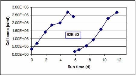
Figure 2. Cell growth curve before and after ball transfer experiment B2B #3
Figures 3 and 4 show the growth of Vero cells on the microcarriers before and after the ball is transferred, with the morphology of the cells on the first, third and fifth days before inoculation, respectively. The top three panels in Figure 4 show the cell growth morphology of the seed culture from the cell factory prior to the ball transfer experiment B2B #3. This is the growth of typical Vero cell seed cultures. Typically more than 90% of Vero cells will be attached to the microcarriers within 4 hours of inoculation and enter the logarithmic growth phase within 24 hours. From the growth of Vero cells on the microcarriers after each ball transfer experiment in Figures 3 and 4, it can be seen that the cells maintained a good growth morphology similar to that of the seed cell culture after the ball transfer experiment.

Figure 3. Growth of Vero cells on microcarriers after ball transfer experiments B2B #1 and B2B #2
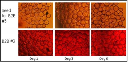
Figure 4. Growth of Vero cells on microcarriers before and after ball transfer experiment B2B #3
High magnification magnification in cell culture bags
We tried to increase the magnification of the ball-to-ball amplification process by a factor of ten in two ways. The first is to culture Vero cells to a cell density of about 4 x 10 6 /ml with a microcarrier density of 6 g/L. After the ball is transferred, a new round of cultivation is started with ten times magnification (culture volume amplification, microcarrier surface area amplification, or both). The second is to culture at a microcarrier density of 3g/L as above, until the cell density is close to 3x10 6
At the time, the ball transfer experiment was carried out, and the magnification of the culture volume and the surface area of ​​the microcarrier was ten times. To compensate for the slow growth that may be caused by the low initial cell density, we tried to culture in a smaller culture volume after the ball was transferred. When the number of cells grows to a certain extent, add new medium to the required volume. Observe the cell growth before and after the ball is transferred.
B2B #4 The first method was used for high-magnification amplification, that is, Vero cells were cultured at a density of 6 g/L of microcarriers to a cell density of 4.3× 10 6 /ml, and the culture volume was 2 liters. Vero cells were digested from the microcarriers using trypsin. One tenth of the cell/microcarrier suspension was inoculated into a new 2 liter culture volume at a microcarrier concentration of 6 g/L such that the culture volume and the microcarrier surface area were magnified ten times. As a parallel comparison test, one tenth of the cell/microcarrier suspension was inoculated into another 2 liter culture volume at a microcarrier concentration of 3 g/L, such that the culture volume was magnified ten times and the microcarrier surface area was magnified five times. The two cultures were carried out simultaneously to observe the growth of the cells.

Figure 5. Cell growth curve before and after B2B #4 in ball-to-ball experiment
Figure 5 shows the cell growth curve before and after the ball transfer experiment B2B #4. Interestingly, the growth rates of the cells in the two parallel cultures after the ball was transferred were basically similar, although the microcarrier density was twice as large. They used 1-2 days more than seed culture to achieve similar cell densities. This should be due to the relatively low initial cell density of the culture after the ball is transferred, but the cell doubling rate is not low. After the ball was transferred, the cell doubling rate was 0.57 d-1 for the first four days of the culture, and the cell doubling rate of the four days before the seed culture was 0.54 d-1.
Figure 6 shows the growth of Vero cells on the microcarriers before and after the ball-to-ball experiment B2B #4. It can be seen that although the number of starting cells is small after the ball is transferred, the cell adherence and survival are still good. The morphology of cell growth was also stable and good in the following days. Although the cells appeared to be unevenly distributed on the microcarriers in the first few days, the cells proliferated normally and quickly spread all the microcarriers in the next few days.
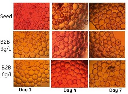
Figure 6. Growth of Vero cells on microcarriers before and after ball transfer experiment B2B #4
B2B #5 uses the second method for high magnification amplification. Vero cells were cultured at a microcarrier density of 3 g/L to a cell density of 3.07 x 10 6 /ml, and the culture volume was 3 liters. Vero cells were digested from the microcarriers using trypsin. One tenth of the cell/microcarrier suspension was inoculated into a new 1.5 liter culture volume at a microcarrier concentration of 6 g/L. When the cell density reached 5x10 6 /ml or more, fresh medium was added to a culture volume of 3 liters. Thus, the magnification of the culture volume and the surface area of ​​the microcarriers was ten times, and the final microcarrier concentration was 3 g/L. In the early stage of culture, the cell density and the microcarrier density are increased, thereby increasing the contact probability of the microcarriers and the cells, and theoretically, the cells are attached to the microcarriers and can grow better. As a parallel comparison test, one tenth of the cell/microcarrier suspension was inoculated into another cell culture bag at a volume of 3 liters and a microcarrier concentration of 3 g/L. The two cultures were carried out simultaneously to observe the growth of the cells.

Figure 7. Cell growth curve before and after ball transfer experiment B2B #5
Figure 7 shows the cell growth curve before and after the above ball transfer experiment B2B #5. Since one of them has a change in culture volume during the culture, the ordinate here uses the total number of cells. The cell growth rate was basically the same before and after the ball was transferred. After the ball is transferred, the cells still maintain vigorous growth vigor. It is also interesting to note that the ball is transferred to a smaller culture volume, which increases cell density and microcarrier density per unit volume, and increases the probability of contact between cells and microcarriers. This method does not significantly increase cell growth rate. . This indicates that the cells grow well at a slightly lower seeding density and do not require a method of lowering the initial culture volume to increase the seeding density.
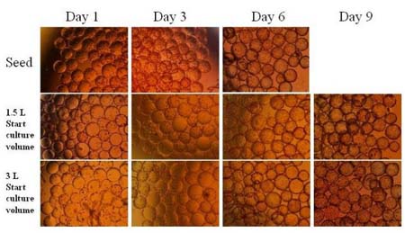
Figure 8. Growth of Vero cells on microcarriers before and after ball transfer experiment B2B #5
Figure 8 shows the growth of Vero cells on the microcarriers before and after the ball-ball experiment B2B #5. The culture time was extended by two days due to the lower initial density of the cells in the culture after the ball was transferred. Therefore, in addition to the cell morphology corresponding to the seed culture in the first, third, and sixth days, we also listed the morphological photographs of the cells on the microcarriers after nine days of culture after the ball was transferred. The cells adhered and grew well after the ball was transferred. We also saw that the initial adherence of cells cultured at initial densities of 1.5 L and 3 L, respectively, and the amount of cells attached, and the subsequent cell growth were essentially the same. This also suggests that the method of increasing cell density and microcarrier density per unit volume, thereby increasing the probability of contact between cells and microcarriers, does not bring much benefit.
discuss
In this part of our work, we performed a ball-to-ball experiment of Vero cells from microcarriers. Cell culture was performed in a WAVETM reactor, and ball-ball experiments were performed in bottles or in WAVETM culture bags. The experimental results show that the ball transfer experiment is quite successful, and it is feasible to expand the microcarrier culture of Vero cells in the WAVETM reactor.
We tried ball-to-ball experiments in bottles (B2B #1) and in WAVETM culture bags (B2B #2 and B2B #3). In the small-scale laboratory, ball-to-ball experiments in bottles have certain advantages over cell culture bags. The former is easier to operate. In a transparent bottle, the settled microcarriers are more easily visible, so the supernatant can be removed more. This allows the microcarrier to be more effectively cleaned. In addition, the bottle can also be kept at a constant temperature by using a water bath, and if it is not too large, it can be shaken more vigorously. This allows the cells to detach from the microcarriers more efficiently. However, if large-scale cultivation is considered, the ball-turning experiment needs to be performed in the WAVETM culture bag because there is a fully enclosed system to better handle larger volumes. From the results of the ball transfer experiment (B2B #2 and B2B #3) in the WAVETM culture bag, the effect of this method is also very good, but more PBS is needed for washing, and the amount of trypsin is also slightly increased.
Trypsin digestion can better separate cells and microcarriers for a longer period of time. However, digestion for too long will also reduce cell viability and affect the effect of cell reattachment. Therefore, trypsin digestion time is best controlled within 40 minutes. In the digestion process, in addition to the digestion time, the sufficient mixing between enzyme dosage, reaction temperature, microcarrier and enzyme is an important factor to ensure the simultaneous digestion of cells on different microcarriers, thus effectively avoiding local excess of enzyme and potential cell damage.
Compared to the stirred tank, the WAVE reactor can have a wider culture range (10-100% working volume) in the same culture bag, providing uniform and effective volume for different reaction volumes of seed amplification and cell digestion. Mixing to achieve in situ digestion of the microcarriers without the need for a specific digestion reactor. The transfer of microcarriers before and after digestion is avoided, and the operation is simple. The uniform and effective mixing is beneficial to precisely control the conditions of the digestion reaction, and the integrity and recovery of the cells are ensured to the greatest extent, which becomes the key to successful amplification of the microcarrier ball.
The results of the ball transfer experiments B2B #4 and #5 suggest that it is completely feasible to perform high-magnification amplification of the ball, and the magnification can be up to ten times. The premise should be to allow the seed culture to reach sufficient cell density to ensure that there are enough The cells are cultured in the next step. Adequate cell seeding density, as well as the ratio of the number of cells to the surface area of ​​the microcarriers, ensure that the cells adhere effectively and rapidly enter the growth phase. Our experiments show that the microcarrier surface area or the change of the culture volume in a small range after the ball is transferred does not have much influence on the adhesion and growth of the cells.
The several ball-turning experiments we have described here are done by transferring a portion of the old microcarriers together with the cells. From the morphology of the cells growing on the microcarriers before and after each ball transfer experiment, the cells can be evenly distributed on the microcarriers. Thus, the transfer of the old microcarriers with the cells does not affect the attachment of the cells to the new microcarriers. If it is not transferred with the old microcarriers, it is necessary to separate the cells from the microcarriers. This is meaning
It takes extra steps and longer process time. These additional steps also require more complex equipment and thus increase the risk of contamination. Since the cells are not distributed unevenly on the microcarriers or more concentrated in the old microcarriers, we do not consider it necessary to separate the cells from the old microcarriers.
in conclusion
The ball-turning experiment in the WAVETM reactor can be successfully carried out by means of cell and microcarrier separation in a bottle or directly in a cell culture bag. And the magnification can easily reach ten times. After transfer, Vero cells maintain similar growth characteristics as seed cells. Vero cells grown on microcarriers were washed with PBS-EDTA pre-warmed at 37 ° C, treated with trypsin pre-warmed at 37 ° C, and trypsin treatment for no more than 40 minutes. It is not necessary to separate the cells from the old microcarriers during the transfer process.
The WAVE microcarrier ball transfer technology provides reliable support for scale-up of microcarrier cell cultures, which can be in situ digested in the same culture bag and then transferred directly to a larger WAVE reactor for re-adhesive expansion. It is not necessary to use a separate digestion reactor, the digestion conditions are easy to control, and the equipment investment cost is low; the aseptic welding machine and the sealing machine combined with the cell culture bag can conveniently realize the aseptic pipeline closed production, which is not only easy to operate but also avoids pollution, and at the same time prevents The operator's exposure to the virus enables safer production operations. Therefore, the large-scale microcarrier cell culture of the WAVE bioreactor can replace the cumbersome transfer bottle and the culture process of several small-scale reactors, and become the development trend of large-scale vaccine production.
Reference article
[1]. History and Characterization of the Vero Cell Line -- A Report prepared by CDR Rebecca Sheets, Ph.D., USPHS CBER/OVRR/DVRPA/VVB for the Vaccines and Related Biological Products Advisory Committee Meeting to be held on May 12, 2000 OPEN SESSION pdf
[2]. Cell culture (Vero) derived whole virus (H5N1) vaccine based on wild-type virus strain induces cross-protective immune responses. Kistner O, Howard MK, Spruth M, Wodal W, Bruohl P, Gerencer M, Crowe BA , Savidis-Dacho H, Livey I, Reiter M and others. 2007, Vaccine 25(32): 6028-6036.
[3]. Flu Vaccine Race Against The Clock. Thayer AM. 2009, Chemical & Engineering News, September 28, 2009, 87(39), p27-33
[4]. A novel mammalian cell (Vero) derived influenza virus vaccine: Development, characterization and industrial scale production. Kistner O, Barrett PN, Mundt W, Reiter M, Schober-Bendixen S, Eder G, Dorner F. 1999. Wiener Klinische Wochenschrift 111(5): 207-214.
[5]. High immunogenic enterovirus 71 strain and its production using serum-free microcarrier Vero cell culture. Liu CC, Lian WC, Butler M, Wu SC. 2007. Vaccine 25(1): 19-24.
We are also able to offer solutions for the meat industry, dairy, for the fish and seafood industry and for other sectors such as catering, gastronomy, bakery and bolleria or sports and diet nutrition.
If
you want to know more about the products in Prepared For Meat Treatment, please click the product details to
view parameters, models, pictures, prices and other information about Prepared for meat treatment.
Whatever you are a group or individual, we will do our best
to provide you with accurate and comprehensive message about Prepared For Meat Treatment!
TGase prepared for meat treatment, TG enzyme for sausage, Transglutaminase enzyme for sausage
Guangdong Kelong Biotechnology Co., Ltd. , https://www.kelongfood.com